How Animal Hospitals Investigate Chest Fluid With Pleural Fluid Analysis
- Pet Care
Elvia Byrd
- 0
- 8 minutes read
You notice your pet is breathing harder, moving less, or standing with their elbows out because lying down seems like too much work. That kind of change gets your attention fast. When a veterinarian providing veterinary services in Hamilton, ON finds fluid in the chest, the concern is immediate because that fluid can press on the lungs and make every breath harder than it should be.
This is where pleural fluid analysis becomes part of the workup. Animal hospitals use it to learn what the fluid is, where it may be coming from, and what disease process is most likely behind it. The sample can point toward infection, bleeding, heart disease, cancer, inflammation, or leakage from the lymphatic system. It does not always give every answer on its own, but it often changes the next step from guessing to treating with purpose.
Pleural fluid analysis helps animal hospitals identify the cause of chest fluid
Chest fluid, also called pleural effusion, is not a diagnosis by itself. It is a sign that something else is happening. Your pet may have fluid because of congestive heart failure, trauma, a ruptured mass, infection, low protein levels, clotting problems, or a condition called chylothorax, where lymphatic fluid collects in the chest. Some pets arrive in distress and need the fluid removed first so they can breathe. Once they are stable enough, the fluid becomes evidence.
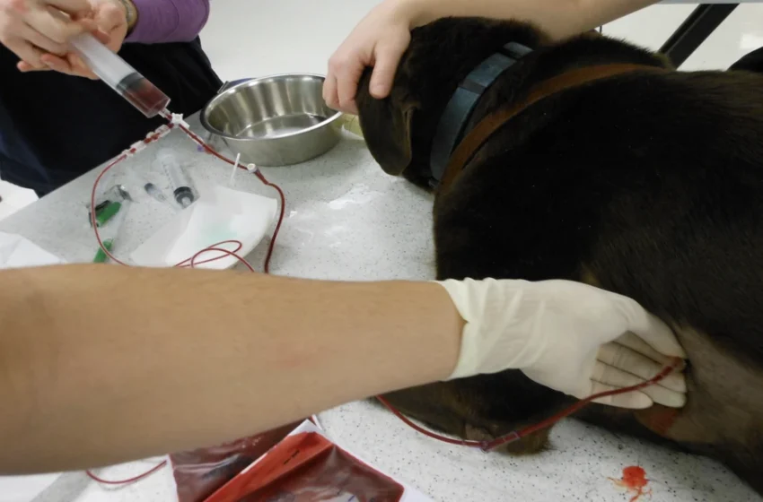
Veterinarians usually collect that sample during thoracocentesis, which means carefully removing fluid from the pleural space with a needle or catheter. For many owners, this moment is frightening because everything feels urgent. The good news is that the procedure often gives relief quickly, and the sample can tell the medical team whether they are dealing with pus, blood, modified transudate, chyle, or another type of effusion.
A chest fluid analysis in pets often includes color, clarity, protein level, cell count, and microscopic review. Under the microscope, a clinical pathologist or veterinarian looks for inflammatory cells, bacteria, abnormal cells, blood contamination, and signs of cancer. Cornell’s guidance on cytology of fluid samples outlines how these samples are assessed and why proper handling matters.
The sample alone does not tell the whole story
This is the part many people do not expect. A fluid sample can be very helpful and still not be the final word. Some cancers do not shed obvious cells into the fluid. Some bloody samples are hard to interpret if the tap itself adds blood. Some infections are visible under the microscope, while others need culture or more advanced testing. That is why animal hospitals rarely rely on one result in isolation.
If your pet has chest fluid, the medical team may pair the analysis with chest radiographs, ultrasound, bloodwork, clotting tests, and sometimes echocardiography. A cat with pleural effusion may need heart evaluation because feline heart disease is a common cause. A dog with a milky sample may need triglyceride testing to confirm chylous effusion. A pet with fever and inflammatory cells may need culture and antibiotics guided by results.
The University of Florida’s clinical pathology laboratory resources show how fluid evaluation fits into a broader diagnostic process. The Minnesota Veterinary Clinical Pathology Lab also provides cytology test protocols that reflect how much sample quality and submission details affect interpretation.
Animal hospital testing for pleural effusion balances urgency and accuracy
When a pet cannot breathe well, speed matters. When the cause is unclear, accuracy matters just as much. That tension is part of why these cases feel so overwhelming. You may hear terms quickly, approve tests quickly, and still feel like the answers are unfolding one layer at a time.
In practice, veterinarians often make decisions in stages. First, relieve pressure on the lungs. Next, examine the fluid and stabilize the patient. Then look for the underlying disease. If the fluid suggests infection, treatment may start right away while cultures are pending. If the sample points toward heart failure, therapy may shift in a very different direction. If cancer is suspected, imaging or biopsy may become the next step. Animal hospital pleural effusion testing is valuable because it narrows that path.
Common pleural fluid findings and what they may suggest
| Fluid finding | What it may suggest | What often comes next |
|---|---|---|
| Clear, low cell count fluid | Transudate linked to low protein or pressure changes | Bloodwork, protein evaluation, heart assessment |
| Cloudy fluid with many inflammatory cells | Infection or sterile inflammation | Cytology review, culture, antibiotics if indicated |
| Frank blood or strongly blood tinged fluid | Bleeding, trauma, clotting disorder, ruptured mass | PCV comparison, imaging, clotting tests |
| Milky white fluid | Chylothorax | Triglyceride testing, imaging, heart evaluation |
| Cells suspicious for neoplasia | Cancer | Imaging, repeat cytology, tissue biopsy |
Three steps you can take right away when chest fluid is suspected
1. Watch breathing, not just energy level. Count your pet’s resting breaths, note any open mouth breathing in cats, and pay attention to posture. Pets with chest fluid may still try to seem normal until they cannot compensate anymore.
2. Ask what the fluid looked like and what the first review showed. You do not need to know every lab term. Ask whether the sample looked like blood, chyle, pus, or a low protein effusion, and whether the team saw bacteria, inflammatory cells, or suspicious cells. That gives you a clearer picture of what the veterinarians are ruling in or out.
3. Ask about the next test that changes treatment. In some cases that is imaging. In others it is culture, heart testing, or repeat sampling. This keeps the plan focused on decisions that matter now, not just a long list of possibilities.
Clear answers often come in stages
If your pet needs pleural fluid analysis, you are probably dealing with a scary day and too many unknowns. The test matters because it turns chest fluid from a symptom into useful clinical information. It helps animal hospital teams sort out what is most likely, what is dangerous right now, and what treatment should happen next.
Stay close to the care plan, ask for the plain language version of the results, and keep the focus on breathing comfort and the underlying cause. An animal hospital can use chest fluid testing, imaging, and clinical pathology together to build the clearest path forward for your pet.
